Corneal tomography interpretation
Tomography derives from the Greek words tomos (to cut) and graphein (to write). The term topography is related to the study of the earth’s surface but is wrongly used in ophthalmology imaging. Who has not seen these corneal tomography images that your doctor used to diagnose your keratoconus? I have always wanted to understand more about these maps of the cornea but was clueless about their meaning.
Brands of imaging systems
There are many different brands of imaging systems: the Orbscan (Bausch & Lomb, Rochester, NY), the Pentacam (Oculus, Inc., Lynnwood, Washington), and the Galilei Dual Scheimpflug Analyzer (Ziemer Ophthalmic Systems AG, Port, Switzerland), but I will explain the data generated by one of the most common ones the Orbscan.
The Orbscan
The Orbscan© shows the front and back shape of the cornea and its thickness. This corneal tomography uses a “best-fit sphere” method to calculate the highs and lows of the surface.
It’s like the sea level that establishes the high of every mountain, city or lake in the world, for instance, New Orleans is 6 feet below sea level and Mexico City is 2200 meters above it.
Tomography colors
These highs and lows are represented with a scale of colors ranging from light green (sea level) to dark red (very high), and below the green from light blues to dark purples for the negative values (below sea level).
The colors in the map represent these altitudes and a difference of usually 10 or 20 microns means the color scale changes. Light colors like green mean near-normal surfaces, yellow and orange colors mean higher areas.
A micron is a size of a millimeter divided by a thousand, just to compare a bacteria can measure 5 microns.
Orbscan views
The Orb scan has four views from the top left and clockwise, (Anterior, Posterior, Thickness, and Keratometric).
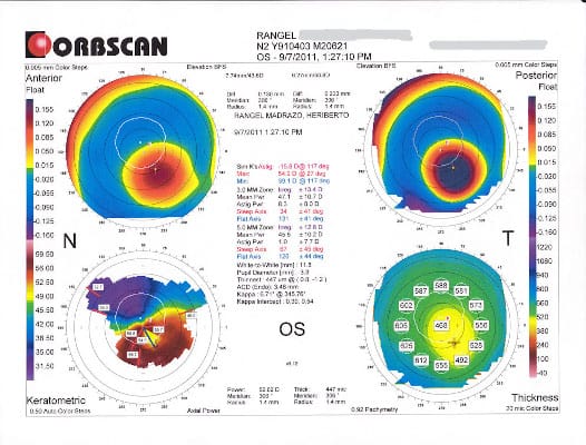
The most useful views for diagnosing are the Posterior, Anterior, and Pachymetry thickness maps, the Posterior float is regarded as the most useful to diagnose developing keratoconus commonly called form fruste keratoconus.
On the image below I have marked some values that you need to compare with normal corneal values to diagnose probable keratoconus. on the top center of the image the two D values from the anterior and posterior maps can be compared with:
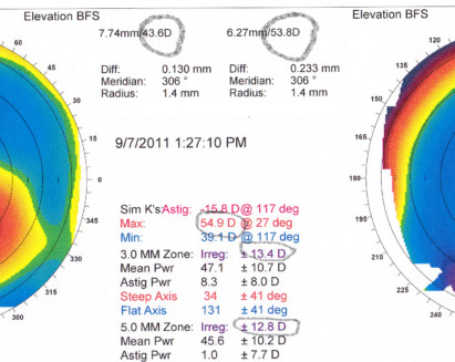
a) Posterior Best fit Sphere less than 52D is normal, more than this abnormal, in this case, 53.8.
b) Sim K’s max keratometry of more than 45.7D is suspect, in this case, 54.9
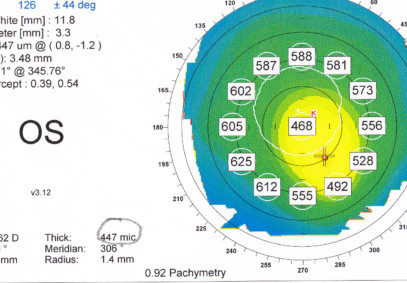
c) On the Pachymetry map on the right and below if we find the thinnest area less than 470 microns it is suspect of thinning due to keratoconus, you can see the lowest value is 447 microns on the center thick value, this is near the apex of the cone.
d) The difference between the central thickness value in this image 468 microns and the thinnest area in this case 447 has to be between 15 and 20 microns, 21 is above and not normal.

Indices
Irregularity at 3mm zone more than 1.5D can suggest Keratoconus. Irregularity at 5mm zone more than 2D to 3D can suggest the same.
Generally speaking, when you see Anterior and Posterior maps with more than three colors this is suspected to be abnormal, in this case blue, orange, yellow, and red.
Corneal Thickness
Elevation maps are more accurate in showing the morphology of the cone and in this case, the elevated areas coincide with the thinnest corneal thickness on the Pachymetry map showing clearly the island formed by the cone protrusion.
The thickness is important since the thickness of the cornea is a value used for qualifying you to treatments like Intacs or crosslinking, usually a minimum of 400 microns thickness.
Biography:
- Belin, Michael W., MD, FACS; Khachikian, Stephen S., MD; Ambrosio Jr., Renato, MD, PhD
“ELEVATION BASED CORNEAL TOMOGRAPHY" © Copyright, 2012 for Jaypee - Highlights Medical Publishers, Inc.
Conclusion
Are you more confused? I hope this helps a bit to understand more about the type of keratoconus you have and its severity. Is important to note that each time you get a corneal tomography the results depend on many variables and in the calibration of the instrument, so different results can occur.
What do you think? if you liked this post leave a comment below.
Good Luck!
Heriberto R.
Hi, I love to improve myself and others. Reach me on hrangelm@gmail.com