keratoconus overview
Keratoconus from Greek: kerato (horn, cornea), and konos cone. Is a condition of the cornea; the cornea is a lens whose primary function is to focus the light rays from our environment the retina in the back of our eye. In the retina, there are receptors called rods and cones that send information about light and color through the optical nerve to the brain.
In the brain, this information is processed and an image is formed, so in reality, we see with our eyes and with our brain. Rods handle the peripheral vision, the vision in low light conditions and cones handle mostly color and detail.
The keratoconus cornea bulges forward and instead of spherical becomes like a cone and therefore the name keratoconus. This bulging produces distorted patterns of light that become heavy myopia and astigmatism (refraction error).
The Cornea
The cornea is one of the most important parts of the eye, if everything is right with your eyes, but your corneas are damaged, you won’t see very well. The cornea is the transparent part of the sclera (the white part), that allows the light rays to enter the eye to be able to see.
You can think of the cornea as the lens of a camera. And it is this important function that people with keratoconus have problems with.
The cornea even though it has no blood vessels except at its margin, is nourished from within the eye and from the outside by the oxygen in the air we breathe.
Cornea layers
- Corneal Epithelium (outer part)
- Bowman’s layer
- Corneal Stroma (90% of the corneal thickness),
- Descemet’s membrane
- Corneal Endothelium (innermost part)
From within the eye, the cornea receives nutrients from the aqueous humor, this is replenished continually and helps maintain the cornea’s convex shape by intraocular pressure.
The aqueous humor is a filtrate of blood and can be thought of as clear blood.1
Even though aqueous humor contains no red blood cells and little protein, it carries oxygen and nutrients to the posterior cornea, lens, and iris, especially ascorbate (vitamin C) which has antioxidant properties.
What causes keratoconus?
It is not known what causes keratoconus: there is a belief that it has a genetic origin because people of the same family with keratoconus have been reported, or that it is secondary to some disease process.
Keratoconus Types of cones
Nipple cone. - small and centrally located.
Oval cone. - large in size between 5 and 6 mm.
Globus cone. - keratoglobus, more than 6mm and hard to fit.
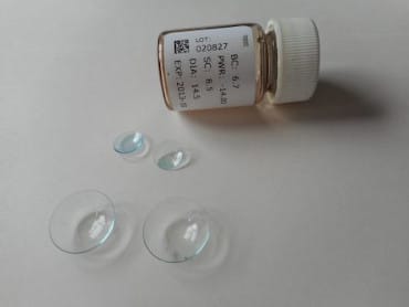
Contact Lenses
Contact lenses work by replacing the irregular surface of the cornea with the more correct shape of the lens so that the light rays are properly focused. In the preliminary stages of the disease, the refraction error can be corrected with glasses or soft contact lenses. Later with increased astigmatism, toric soft contact lenses work for a while, but gradually rigid permeable gas contact lenses (RGP’s) are necessary to correct the distorted cornea and obtain acceptable vision.
1 David R. Whikehart. Biochemistry of the eye. 2nd ed. Butterworth-Heinemann, 2003:9-10.
Good Luck.